
Key facts
- Bond type
- Amide bond, -CO-NH-
- Formed by
- Condensation: loss of one H2O (18.02 g/mol)
- Bonds in a linear chain
- n amino acids give n - 1 peptide bonds
- Geometry
- Planar, with partial double-bond character
- Usual configuration
- Trans; cis is uncommon except before proline
- Broken by
- Hydrolysis (acid, base or protease enzymes)
Definition: What Is a Peptide Bond?
A peptide bond is a covalent amide linkage between the carbonyl carbon of one amino acid and the alpha-amino nitrogen of the next [1]. Chemically, it's the same functional group you find in nylon and plenty of synthetic amides. Biochemists give it its own name because it is the repeating link in every peptide and protein chain.
The atoms fall into a repeating backbone pattern: nitrogen, alpha carbon, carbonyl carbon, then the next nitrogen. Side chains hang off the alpha carbons, but the peptide bonds look the same from one residue to the next. So in a diagram the backbone appears uniform, and it's the side chains that give each peptide its character.
How Peptide Bonds Form
It forms by condensation. The carboxyl group of one amino acid reacts with the amino group of another, and a molecule of water comes off.
Stir two amino acids into water and nothing useful happens. In water the reaction runs thermodynamically uphill, so cells and chemists alike activate the carboxyl group first. Cells load the amino acid onto a transfer RNA, and the ribosome's peptidyl transferase center lines up the reacting groups so the bond can form; crystal structures show that center is made of RNA, not protein [5][6]. In the lab, coupling reagents turn the carboxyl group into a reactive ester or a similar intermediate. Peptide synthesis explained covers that chemistry.
A worked example: counting bonds and water
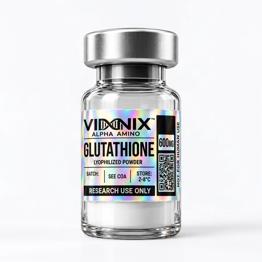
Glutathione has three residues, so it has two linkages, and making it releases two water molecules. One of those links is odd. Glutamate joins cysteine through its side-chain gamma carboxyl group, forming an amide that is chemically similar to a standard peptide bond but sits outside the regular alpha backbone. The Cys-Gly link is an ordinary peptide bond. Hence the way glutathione is written: gamma-Glu-Cys-Gly.
Peptide Bond Structure: Resonance and Planarity
Electrons are shared across the carbonyl oxygen, the carbonyl carbon and the nitrogen. That sharing gives the C-N bond partial double-bond character, and it's why the peptide bond is planar.
Linus Pauling and Robert Corey built this geometry into their 1951 models of protein structure. Their planar amide group had a C-N distance of about 1.32 angstroms, shorter than the roughly 1.47 angstroms of a typical C-N single bond [2]. Six atoms around the bond lie in one plane, usually called the peptide plane: the two alpha carbons, the carbonyl C and O, and the N with its H.
- Restricted rotation. Partial double-bond character stops the peptide bond itself from rotating freely. The backbone gets its flexibility from the two single bonds on either side of each alpha carbon, described by the phi and psi angles.
- Polarity. The carbonyl oxygen carries a partial negative charge and the N-H hydrogen a partial positive one, so backbone groups act as hydrogen-bond acceptors and donors. Those hydrogen bonds hold alpha helices and beta sheets together [1][2].
- Little basicity. Resonance ties up the nitrogen's lone pair, so the amide nitrogen isn't basic the way a free amino group is.
Cis and trans peptide bonds
A rigid bond can sit in two configurations. In trans, the two alpha carbons are on opposite sides of the bond; in cis, they're on the same side. Trans wins by a wide margin because it keeps neighboring side chains out of each other's way. One survey of protein crystal structures found cis peptide bonds in roughly 5% of bonds preceding proline, against only about 0.03% of all other peptide bonds [3]. Proline breaks the pattern because its side chain loops back to its own nitrogen, which brings the cis and trans forms closer in energy.
Peptide Chains
Link enough amino acids with peptide bonds and you get chains, which are named by length.
| Name | Residues | Peptide bonds (linear) | Example |
|---|---|---|---|
| Dipeptide | 2 | 1 | Carnosine (beta-Ala-His) |
| Tripeptide | 3 | 2 | GHK, KPV, glutathione |
| Oligopeptide | about 2 to 20 | 1 to about 19 | BPC-157 (15), MOTS-c (16) |
| Polypeptide | longer chains | n - 1 | Tesamorelin (44 plus modification) |
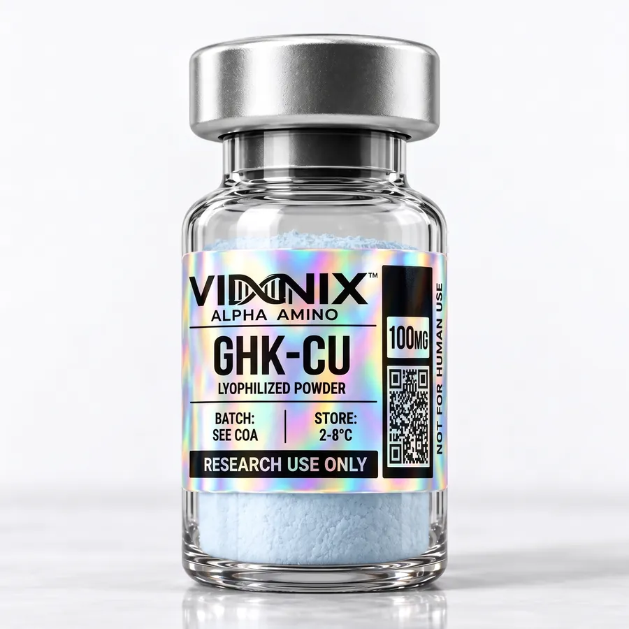
In the Vinnix catalog, GHK-Cu and KPV are tripeptides, and BPC-157 has 15 residues. Cyclic peptides are the exception: once the two ends are joined, or a ring is closed through side chains, the bond count no longer follows the simple n minus 1 rule.
Directionality
Every linear chain has a direction. One end carries a free amino group (the N-terminus), the other a free carboxyl group (the C-terminus), and sequences are written N to C.
That direction comes straight from the bond. Every peptide bond points the same way along the backbone, carbonyl to amide nitrogen, so reading the chain backwards describes a different molecule. Ribosomes build chains N to C as well [6]. Chemical solid-phase synthesis usually goes the opposite way, adding residues from the C-terminus toward the N-terminus. For how this shows up in written sequences, see N-terminus and C-terminus notation.
Hydrolysis: How Peptide Bonds Break
Hydrolysis breaks it. That's formation run in reverse: water is added back across the C-N bond.
With no catalyst, the reaction is extremely slow at neutral pH and room temperature. Kinetic measurements in neutral solution put the half-life of an unactivated peptide bond at 25 degrees C on the order of years [4]. Protease enzymes speed the same reaction up enormously, which is why proteases, not plain water, do most of the peptide breakdown in biological samples.
Labs also hydrolyze peptides on purpose. Amino-acid analysis uses complete hydrolysis with strong acid at high temperature, and it supports net peptide content measurements. Partial hydrolysis is one of several degradation routes analysts watch for, alongside oxidation of methionine and cysteine and deamidation of asparagine. Our peptide purity guide shows how related impurities appear on an HPLC chromatogram.
Why Peptide Bonds Matter
Every peptide and protein is built on a backbone of these bonds, and their geometry limits how chains can fold.
- They fix the order of residues. Sequence and identity rest physically on them.
- Their planarity and hydrogen-bonding pattern sit underneath secondary structures such as helices and sheets [1][2].
- Without enzymes around, they're stable enough that peptides can be synthesized, purified and analyzed without falling apart.
- Forming them is the core reaction of chemical peptide synthesis, so coupling efficiency at each bond shapes the impurity profile of the final material.
Terms such as residue, amide and hydrolysis are defined in the peptide glossary.
FAQFrequently asked questions
What is a peptide bond?
It's the amide bond (-CO-NH-) joining the carboxyl carbon of one amino acid to the amino nitrogen of the next, and the repeating link in every peptide and protein backbone. Resonance makes each bond planar and rigid. In a linear chain, n amino acids are connected by n minus 1 peptide bonds.
How is a peptide bond formed?
By condensation. The carboxyl group of one amino acid joins the amino group of another, and one water molecule is released. First, though, the carboxyl group has to be activated. Cells handle that with transfer RNA and the ribosome; chemists use coupling reagents during solid-phase peptide synthesis.
Why is the peptide bond planar?
The nitrogen shares its lone pair with the neighboring carbonyl group, which gives the C-N bond partial double-bond character. That resonance holds the carbonyl carbon, oxygen, nitrogen, amide hydrogen and the two flanking alpha carbons in a single plane, and it stops the bond itself from rotating freely.
What is the difference between cis and trans peptide bonds?
In a trans peptide bond, the two alpha carbons sit on opposite sides of the C-N bond. In a cis bond, they're on the same side. Trans is strongly favored. Structural surveys find cis bonds in about 5% of bonds that come before proline, but in only about 0.03% of other peptide bonds.
Is a peptide bond the same as an amide bond?
Chemically, yes. A peptide bond is an amide bond formed specifically between the alpha-carboxyl group of one amino acid and the alpha-amino group of another. Amide links that involve side chains, like the gamma-glutamyl bond in glutathione, are sometimes called isopeptide bonds to set them apart from regular backbone peptide bonds.
How are peptide bonds broken?
By hydrolysis, which adds water back across the C-N bond. Without a catalyst that's very slow at neutral pH, with half-lives on the order of years. Protease enzymes, strong acid or strong base speed it up dramatically, and acid hydrolysis is used deliberately in amino-acid analysis.
REFScientific references
-
Alberts B, Johnson A, Lewis J, Raff M, Roberts K, Walter P. Molecular Biology of the Cell. 4th ed. New York: Garland Science; 2002. The Shape and Structure of Proteins. NCBI Bookshelf NBK26830. Source
textbook (NCBI Bookshelf) -
Pauling L, Corey RB, Branson HR. The structure of proteins; two hydrogen-bonded helical configurations of the polypeptide chain. Proc Natl Acad Sci U S A. 1951;37(4):205-211. PubMed 14816373
structural study -
Jabs A, Weiss MS, Hilgenfeld R. Non-proline cis peptide bonds in proteins. J Mol Biol. 1999;286(1):291-304. PubMed 9931267
structural database analysis -
Radzicka A, Wolfenden R. Rates of uncatalyzed peptide bond hydrolysis in neutral solution and the transition state affinities of proteases. J Am Chem Soc. 1996;118(26):6105-6109. doi:10.1021/ja954077c (not indexed in PubMed; verified via Crossref).
in vitro kinetic study -
Nissen P, Hansen J, Ban N, Moore PB, Steitz TA. The structural basis of ribosome activity in peptide bond synthesis. Science. 2000;289(5481):920-930. PubMed 10937990
structural study (X-ray crystallography) -
Alberts B, Johnson A, Lewis J, Raff M, Roberts K, Walter P. Molecular Biology of the Cell. 4th ed. New York: Garland Science; 2002. From RNA to Protein. NCBI Bookshelf NBK26829. Source
textbook (NCBI Bookshelf)
 Research Library: Peptide Fundamentals
 Research Library: Peptide Fundamentals
 Peptides
 Peptides