Key facts
- Compound class
- Dinucleotide coenzyme (not a peptide)
- Redox partner
- NADH (reduced form)
- Main consuming enzymes
- Sirtuins, PARPs, CD38
- Common precursors studied
- Nicotinamide, NR, NMN, nicotinic acid, tryptophan
- Measurement methods
- Enzymatic cycling assay, LC-MS
- Absorbance
- NAD+ and NADH at 260 nm; only NADH at 340 nm
- pH stability
- NAD+ labile in alkali; NADH labile in acid
What is NAD+ and why is it not a peptide?
NAD+ is two nucleotides joined through their phosphates. One carries adenine, the other carries nicotinamide, and the business end is the nicotinamide ring. There are no amino acids and no peptide bonds, so it sits in a peptide catalog only because it turns up in the same cell metabolism experiments. Its formula, mass and identifiers are on the NAD+ product page.
Cells use it in two very different ways. As a redox coenzyme it is borrowed and returned. As a substrate for signaling enzymes it is broken apart and has to be rebuilt. That second role is why so much research tracks the size of the NAD+ pool rather than just its redox state [1].
What is the difference between NAD+ and NADH?
NAD+ accepts a hydride ion (a proton plus two electrons) at carbon 4 of the nicotinamide ring and becomes NADH. Dehydrogenases in glycolysis and the citric acid cycle run that reaction, and complex I of the mitochondrial respiratory chain runs it backward, handing the electrons on [1].
The two forms look different to a spectrophotometer, which is the basis of many classic assays. Both absorb at 260 nm through the adenine ring. Only NADH absorbs at 340 nm (molar absorptivity about 6,220 per molar per centimeter) and fluoresces when excited there. Watching the 340 nm signal rise or fall is still the standard way to follow a dehydrogenase reaction.
A related molecule, NADP+, carries an extra phosphate on the adenosine ribose. It is handled by a largely separate set of enzymes, and a good assay has to tell the four species, NAD+, NADH, NADP+ and NADPH, apart [4].
Which enzymes consume NAD+?
Three enzyme families cleave NAD+ instead of just reducing it. All of them release nicotinamide, which the cell recycles back into NAD+ through the salvage pathway [1][2].
| Enzyme family | Reaction | What labs studied |
|---|---|---|
| Sirtuins (SIRT1 to SIRT7) | Remove acyl groups from proteins, using NAD+ and releasing nicotinamide | Protein deacetylation, mitochondrial enzyme regulation |
| PARPs (e.g. PARP1) | Build ADP-ribose chains on proteins | DNA damage signaling and repair |
| CD38 | Hydrolyzes NAD+ (NADase); also degrades NMN | NAD+ decline in aging mouse tissues |
CD38 drew particular attention after a 2016 mouse study from Camacho-Pereira and colleagues. They reported that CD38 expression and activity rose with age in mouse tissues, that mice lacking CD38 did not show the age-related NAD+ drop, and that CD38 was the main enzyme degrading NMN in vivo [3]. Those were mouse results, and the authors linked part of the effect to SIRT3 activity in mitochondria [3].
What are NR, NMN and nicotinamide?
Cells build NAD+ along three routes: from tryptophan (de novo), from nicotinic acid (the Preiss-Handler pathway) and from recycled nicotinamide (salvage). Nicotinamide riboside (NR) and nicotinamide mononucleotide (NMN) feed into the salvage route close to its end [2].
| Precursor | Entry point | Key enzyme |
|---|---|---|
| Tryptophan | De novo pathway via kynurenine | Several steps to quinolinic acid |
| Nicotinic acid | Preiss-Handler pathway | NAPRT |
| Nicotinamide | Salvage pathway | NAMPT, converts it to NMN |
| Nicotinamide riboside (NR) | Phosphorylated to NMN | NR kinases (NRK1, NRK2) |
| Nicotinamide mononucleotide (NMN) | Adenylated to NAD+ | NMNAT1 to NMNAT3 |
Yoshino, Baur and Imai reviewed NR and NMN in 2018 and stressed that their pharmacokinetics and metabolic fates were still being worked out. How each one reaches the inside of a cell, and in which tissues, depends on nucleotidases, kinases and presumptive transporters that differ between tissues [2]. Whether NMN crosses membranes intact or is first converted to NR outside the cell has been debated.
How is NAD+ measured in the lab?
Two families of methods dominate, and Kanamori and colleagues published a method that runs both side by side on the same tissue and cell extracts [5].
Enzymatic cycling assays
A cycling assay amplifies a tiny amount of NAD+ by running it around a loop. Alcohol dehydrogenase reduces NAD+ to NADH using ethanol, then a second enzyme such as diaphorase reoxidizes the NADH while reducing a dye. Each pass through the loop adds colored or fluorescent product, so the signal grows with time in proportion to the starting NAD+ [5]. The method is cheap and sensitive, but it reports total NAD(H) unless the sample is split and treated to destroy one form first.
LC-MS
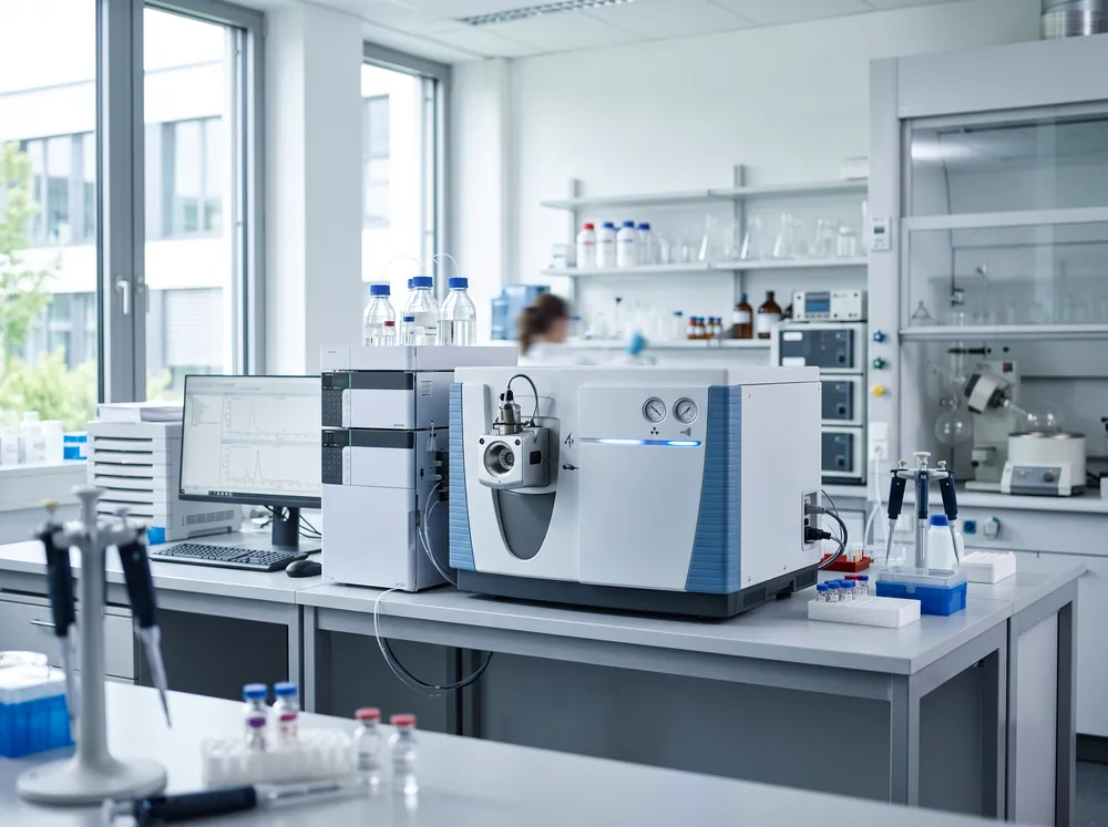
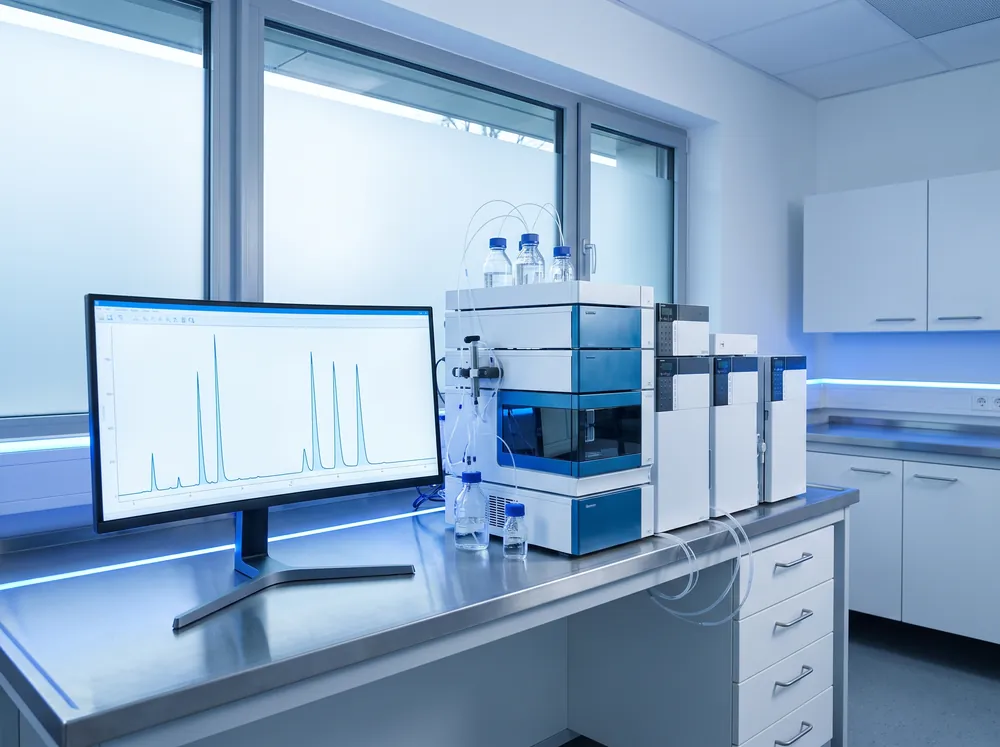
LC-MS separates the molecules by chromatography and then identifies each by mass. One run can resolve NAD+, NADH, NADP+, NADPH, NMN, NR and nicotinamide, and stable-isotope internal standards correct for losses. The trade-off is instrument time and cost (the broader comparison is in HPLC vs. mass spectrometry).
| Feature | Enzymatic cycling | LC-MS |
|---|---|---|
| Specificity | Depends on enzyme and sample split | High, by retention time and mass |
| Species per run | One form, or total NAD(H) | Many metabolites at once |
| Equipment | Plate reader | LC-MS system |
| Main error source | Interfering enzymes, incomplete form destruction | Interconversion and degradation during extraction |
The extraction problem
Both methods are only as good as the extract. Lu and colleagues at Princeton compared seven quenching and extraction solvents and found that NAD+ and NADH interconverted substantially in most of them [4]. A cold mixture of 40:40:20 acetonitrile, methanol and water with 0.1 M formic acid gave the least interconversion and the best recovery, provided the extract was neutralized promptly to stop acid-catalyzed breakdown [4]. They tracked the artifact by growing cells on 13C-glucose and spiking unlabeled standards into the solvent [4].
Why is NAD+ unstable in solution?
The weak point is the bond between nicotinamide and its ribose. In 1961 Lowry, Passonneau and Rock set out the classic stability rules for pyridine nucleotides: the oxidized forms are destroyed by alkali and survive mild acid, while the reduced forms are destroyed by acid and survive alkali [6]. Cycling assays still use that asymmetry, heating split samples in acid or base to remove one form before measuring the other.
- pH. NAD+ solutions keep best near neutral to slightly acidic pH. Alkaline buffers cleave off nicotinamide.
- Temperature. Breakdown speeds up with heat. Keep working solutions on ice and frozen stocks frozen.
- Light. NADH absorbs strongly in the near UV and is the more light-sensitive form. Amber tubes or foil are a sensible default for both.
- Enzymes. Biological samples contain NADases and nucleotidases. Quench them quickly, or the sample loses NAD+ before it reaches the assay.
How should NAD+ be stored and handled at the bench?
General guidance for dry research materials is in our storage guide, and the background on freeze-dried solids is in what is lyophilization. For NAD+ specifically:
- Store the dry powder sealed, cold, dark and with desiccant. It is hygroscopic, so warm the vial to room temperature before opening.
- Dissolve in water or a near-neutral buffer, not an alkaline one, and keep the solution on ice while working.
- Check concentration by absorbance at 260 nm (molar absorptivity about 18,000 per molar per centimeter) rather than trusting weighed mass.
- Check the 340 nm reading. A fresh NAD+ solution should show almost none; a rising 340 nm signal means NADH has appeared.
- Split stock into single-use aliquots, freeze them, and avoid repeated freeze-thaw cycles.
- Record lot number, preparation date and pH on every tube.
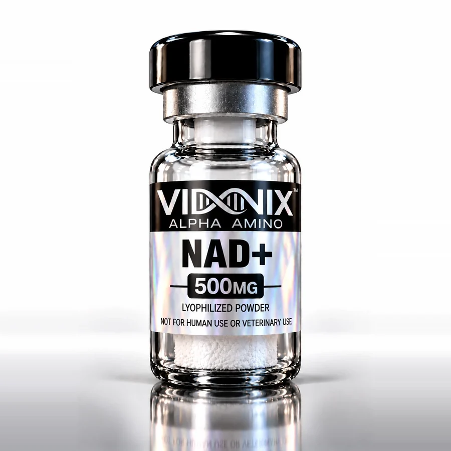
Vinnix supplies NAD+ for laboratory research. It is not for human or veterinary use, and nothing here describes use in people or animals.
What should a lab ask for when sourcing NAD+?
For any supplier, ask for the batch number, HPLC purity with the chromatogram and method, and mass spectrometry showing the observed mass against the expected value. Ask whether NADH, nicotinamide and ADP-ribose were looked for as impurities, since they are the expected breakdown products, and whether the result is reported as the free acid or a salt. Our guide to what a COA should contain lists the fields to check.
FAQFrequently asked questions
Is NAD+ a peptide?
No. NAD+ is a dinucleotide coenzyme built from two nucleotides joined through a pyrophosphate bridge. It contains no amino acids or peptide bonds, and is grouped with peptides only because both appear in the same cell and metabolism experiments.
How do labs measure NAD+?
Most use either an enzymatic cycling assay, which amplifies NAD+ through a dehydrogenase loop and reads a colored or fluorescent dye, or LC-MS, which separates and identifies NAD+ and related metabolites by mass. Both depend on an extraction that stops NAD+ and NADH from interconverting.
Why does NAD+ degrade in solution?
The bond between nicotinamide and its ribose is the weak point. Oxidized NAD+ is destroyed in alkaline solution and is more stable in mild acid, while NADH behaves the opposite way. Heat, light and contaminating enzymes speed up the loss.
What is CD38 and why is it studied with NAD+?
CD38 is an enzyme that hydrolyzes NAD+ and also breaks down NMN. A 2016 mouse study reported that CD38 activity rose with age and that mice lacking CD38 did not show the usual age-related fall in tissue NAD+.
What is the difference between NR and NMN?
Both are NAD+ precursors in the salvage pathway. Nicotinamide riboside is a nucleoside that NR kinases phosphorylate to NMN, and NMN is a nucleotide that NMNAT enzymes convert to NAD+. How each enters cells in different tissues is still being studied.
How can I tell if an NAD+ solution has degraded?
Measure absorbance at 260 and 340 nm. Concentration comes from the 260 nm reading, and a fresh NAD+ solution should show almost no 340 nm absorbance. A rising 340 nm signal points to NADH, and HPLC can show nicotinamide or ADP-ribose from breakdown.
REFScientific references
-
Covarrubias AJ, Perrone R, Grozio A, Verdin E. NAD+ metabolism and its roles in cellular processes during ageing. Nat Rev Mol Cell Biol. 2021;22(2):119-141. PubMed 33353981
review -
Yoshino J, Baur JA, Imai SI. NAD+ intermediates: the biology and therapeutic potential of NMN and NR. Cell Metab. 2018;27(3):513-528. PubMed 29249689
review -
Camacho-Pereira J, Tarrago MG, Chini CCS, Nin V, Escande C, Warner GM, et al. CD38 dictates age-related NAD decline and mitochondrial dysfunction through an SIRT3-dependent mechanism. Cell Metab. 2016;23(6):1127-1139. PubMed 27304511
animal study -
Lu W, Wang L, Chen L, Hui S, Rabinowitz JD. Extraction and quantitation of nicotinamide adenine dinucleotide redox cofactors. Antioxid Redox Signal. 2018;28(3):167-179. PubMed 28497978
in vitro (analytical method) -
Kanamori KS, de Oliveira GC, Auxiliadora-Martins M, Schoon RA, Reid JM, Chini EN. Two different methods of quantification of oxidized nicotinamide adenine dinucleotide (NAD+) and reduced nicotinamide adenine dinucleotide (NADH) intracellular levels: enzymatic coupled cycling assay and ultra-performance liquid chromatography (UPLC)-mass spectrometry. Bio Protoc. 2018;8(14):e2937. PubMed 30101155
analytical method -
Lowry OH, Passonneau JV, Rock MK. The stability of pyridine nucleotides. J Biol Chem. 1961;236:2756-2759. PubMed 14466980
in vitro (chemical stability) -
McReynolds MR, Chellappa K, Baur JA. Age-related NAD+ decline. Exp Gerontol. 2020;134:110888. PubMed 32097708
review
 Research supplies
 Laboratory Practice
 Research Library